Beyond "Normal": How Quantitative Neuroimaging Informs Expert Evaluation of Mild TBI
In mild traumatic brain injury litigation, one of the most persistent challenges is the gap between what a patient experiences and what standard imaging shows. When a routine MRI or CT scan returns "within normal limits," it is not the end of the neurological inquiry. For a qualified neuroimaging expert, it is frequently the beginning of a more nuanced evaluation.
What Standard Imaging Misses, and Why
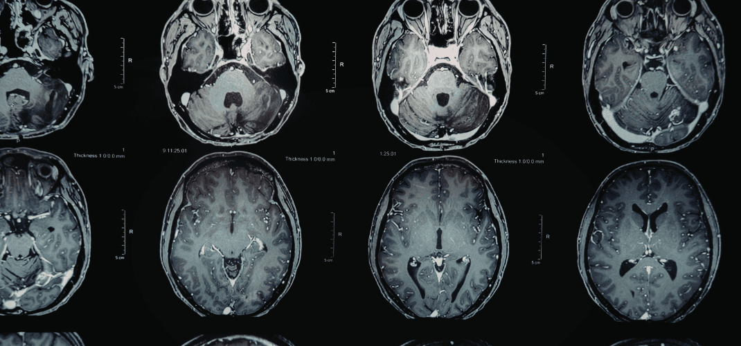
Conventional MRI and CT were not designed to evaluate the integrity of white matter microstructure—the vast network of axonal pathways that connects regions of the brain and underlies cognitive function.
- The Limitation: A scan that is "normal" by standard clinical read has not ruled out microstructural injury.
- The Reality: It simply used a tool not calibrated to detect it.
Diffusion Tensor Imaging (DTI) operates differently. Rather than producing a conventional anatomical image, DTI measures the movement of water molecules through the white matter tracts.
Understanding Diffusion and Fractional Anisotropy (FA)
In healthy, intact white matter, water diffuses in a highly organized, directional manner. When white matter integrity is disrupted—whether through axonal injury or other pathology—that organized directionality breaks down.
Fractional Anisotropy (FA) is the primary metric used to quantify this effect.
- High FA Values: Reflect organized, directional diffusion.
- Low FA Values: Reflect greater isotropy (randomness).
Statistically significant reductions in FA, measured against a validated normative reference population, provide a quantifiable, objective indicator that white matter microstructure deviates from what is expected in a healthy individual.
What a Quantitative Finding Can and Cannot Tell Us
It is important to be precise about what an FA value represents. Reduced FA in a bundle of white matter tracts indicates a measurable deviation from normative expectations, but it does not, on its own, establish a diagnosis.
A qualified neuroimaging expert brings the clinical judgment to evaluate the data in context by:
- Pattern Analysis: Examining the distribution of findings across white matter tracts.
- Biomechanical Consistency: Assessing if findings match the reported injury forces.
- Alternative Explanations: Distinguishing traumatic findings from non-traumatic pathology.
The Role of Convergent Evidence
The evidentiary value of DTI findings is substantially strengthened by their integration with other clinical data. A rigorous expert analysis may draw on:
- Documented symptom history.
- Neuropsychological testing.
- Pre-existing and alternative conditions.
- Additional history, testing, and records.
When these data streams converge, an appropriately qualified expert is in a position to offer an opinion grounded in integration, not in any single metric in isolation.
Move Your Case Forward
MINDSET's Case Consultation service brings qualified neuroimaging and licensed experts into this evaluative process to help you bridge the gap between "normal" imaging and the objective reality of your client's injury.